Revvity Sites Globally
Select your location.
*e-commerce not available for this region.

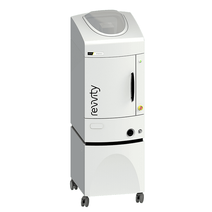
IVIS Spectrum 2 In Vivo Imaging System
For research use only. Not for use in diagnostic procedures.
Part #:
CLS158738
Imaging Modality:
2D and 3D Bioluminescence, 2D and 3D Fluorescence


IVIS Spectrum 2 In Vivo Imaging System


IVIS Spectrum 2 In Vivo Imaging System
IVIS Spectrum 2 In Vivo Imaging System
Supporting products you might need
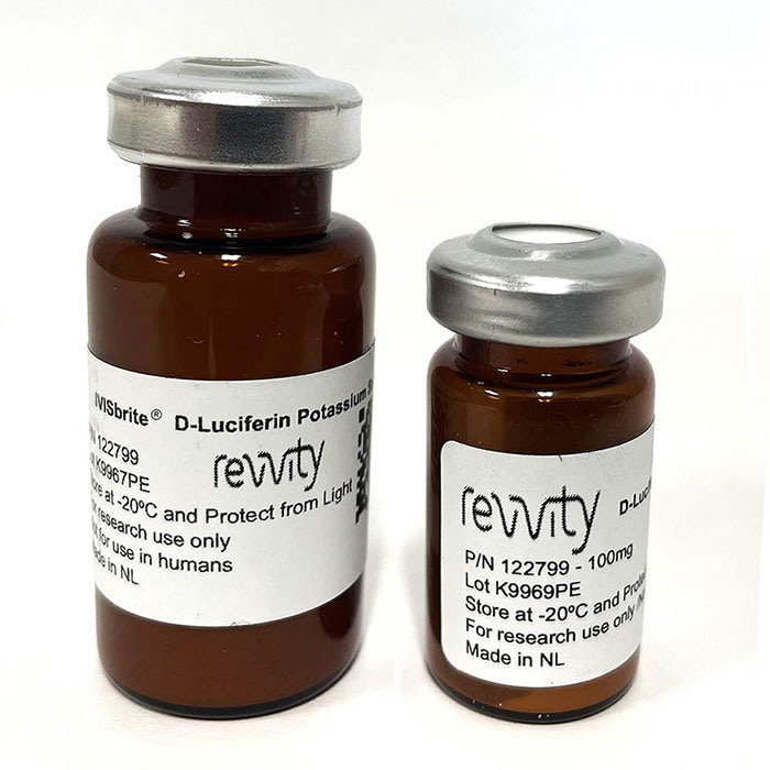
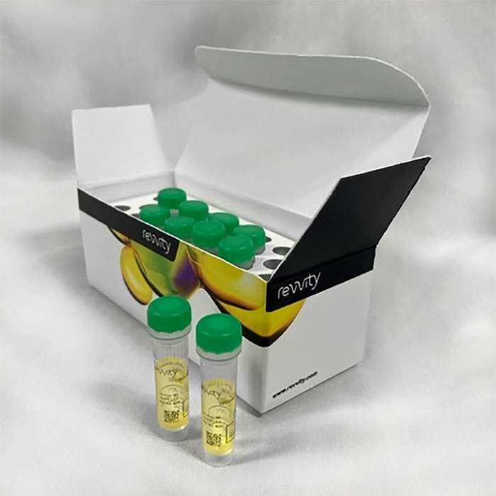
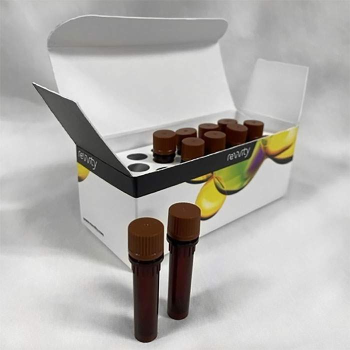
Need D-Luciferin substrate in powder or ready-to-use injectable format?
Product information
Overview
Building upon the IVIS Spectrum in vivo optical system with proven 2D bioluminescence and 2D fluorescence imaging and 3D optical tomography capabilities combined in a single system, the IVIS Spectrum 2 is our next generation in optical imaging. This advanced imaging system incorporates a CCD camera with eXcelon® coating that enables detection of more signal at higher efficiency across a broader spectrum of wavelengths. With exclusivity to this innovative camera for in vivo imaging, the IVIS Spectrum 2 preclinical optical imaging system delivers the sensitivity you demand for non-invasive longitudinal imaging to better understand early disease-related biological changes, track disease progression, and help guide the drug development process.
Additional product information
Features and benefits
![]()
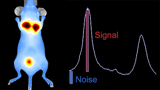
High sensitivity
Exclusivity to patented CCD camera with eXcelon coating for high sensitivity imaging

Rapid imaging
Fast data acquisition allows quick visualization of images in real-time

High throughput
Standard 5 mice configuration or up to 10 mice capacity using optional manifold

Trans-illumination
Imaging below the specimen for sensitive detection and quantification of deep fluorescent sources

Epi-illumination
Imaging above the specimen ideal for high throughput workflow

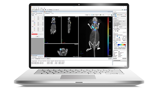
Co-registration
Seamless co-registration of optical data with other modalities, e.g., CT, MRI, SPECT, PET, Ultrasound

Spectral unmixing
Remove autofluorescence & easily identify, separate, and quantify multiplexed fluorescent signals

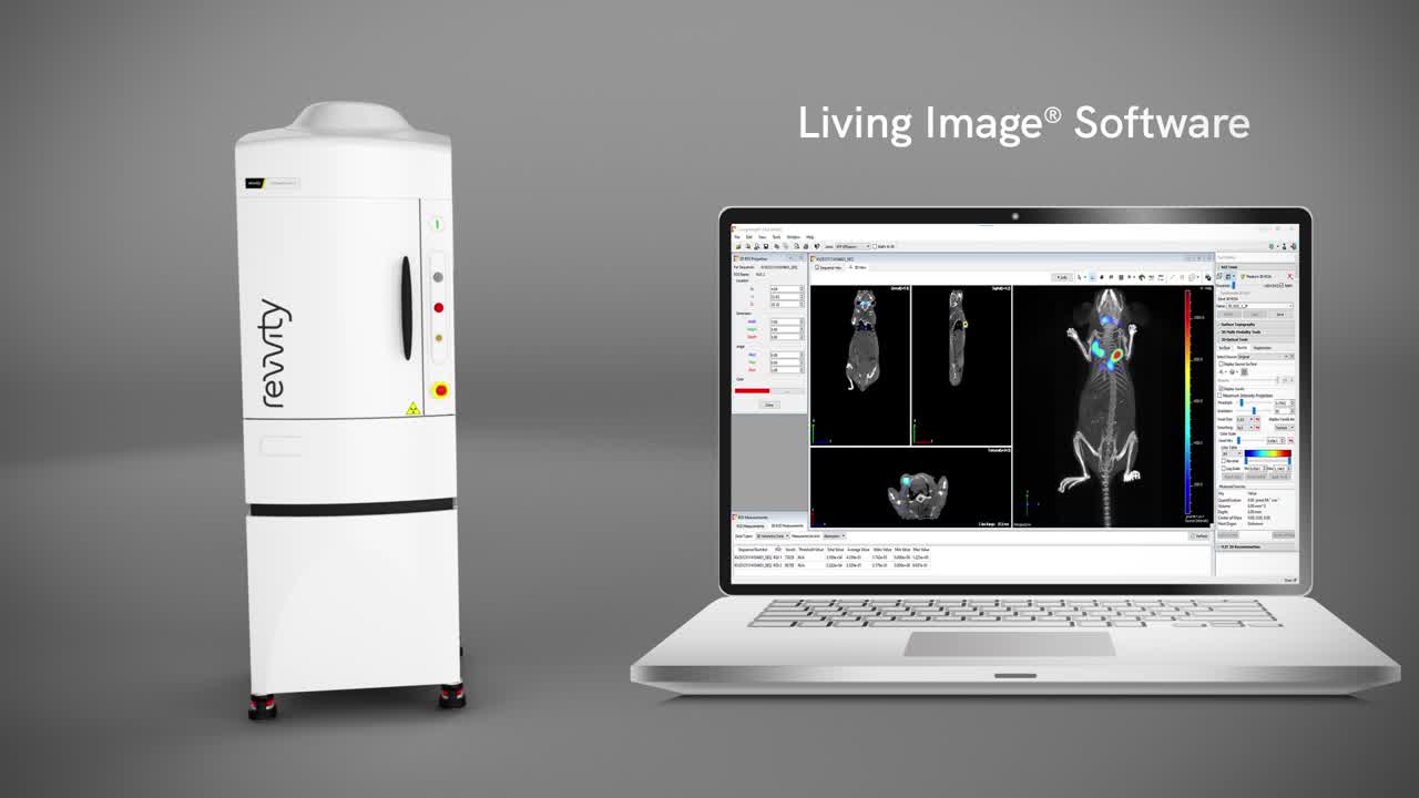
Analysis software
Broadly adopted, easy to use, and intuitive, Living Image® visualization and analysis software
Complimentary Living Image™ software licenses are provided with the IVIS systems and upon request.
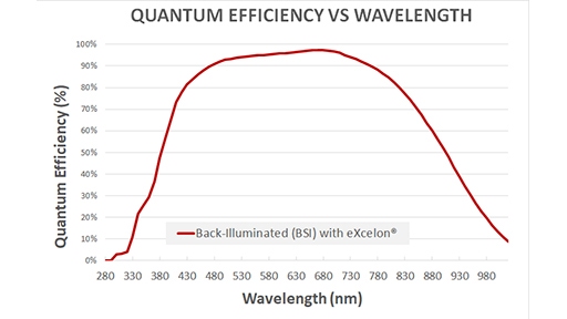
High-performance CCD camera
The camera and coating facilitate detection of more signal at higher efficiency across a broader spectrum of wavelengths throughout the visible and NIR spectrum - giving you:
- Improved signal-to-noise ratio for both bioluminescent and fluorescent signals
- Increased bandwidth to encompass a wider range of NIR fluorescent probes
Camera highlights
- Patented eXcelon® coating
- Back-illuminated, thermoelectrically cooled (-90°C) CCD
- High quantum efficiency (peak >95%)
- 2048 x 2048 imaging pixels with 13.5 micron pixel size
- Low read noise
Bring your in vivo images to life with Living Image software
Broadly adopted imaging software that sets the industry standard for ease of use and flexibility
- Comprehensive set of tools for 2D and 3D data analysis
- One click 3D reconstructions
- Spectral unmixing algorithms to easily obtain and separate simultaneous fluorescent readouts or remove unwanted autofluorescence
- Co-register optical imaging with other modalities (e.g., CT, MRI, SPECT, PET)
- Auto settings for easy image acquisition
- Batch processing analysis tools
- Generation of animated movies and publication ready figures
- Flexible remote review for convenient offline analysis of data sets
- Included in IVIS purchase
Which IVIS system is best for your research?
| IVIS Spectrum 2 | IVIS SpectrumCT 2 | IVIS Lumina S5 | IVIS Lumina X5 | IVIS Lumina III | IVIS Lumina LT | IVIS Lumina XRMS | |
|---|---|---|---|---|---|---|---|
| Capacity | Up to 10 mice* | Up to 10 mice* | Up to 10 mice* | Up to 10 mice* | Up to 5 mice** | Up to 5 mice** | Up to 3 mice |
| Benchtop format | ✔ | ✔ | ✔ | ✔ | ✔ | ||
| 2D Bioluminescence / Fluorescence | ✔/✔ | ✔/✔ | ✔/✔ | ✔/✔ | ✔/✔ | ✔/✔ | ✔/✔ |
| 3D Bioluminescence / Fluorescence | ✔/✔ | ✔/✔ | |||||
| Enhanced Fluorescence capabilities | ✔ | ✔ | ✔ | ✔ | ✔ | ✔ | |
| Integrated standard x-ray | ✔ | ||||||
| Integrated high resolution x-ray | ✔ | ||||||
| Integrated CT | ✔ | ||||||
| Optical FOV (cm) (Nominal) | 4-22.5 | 4-22.5 | 10-22.5 | 10-22.5 | 5-12(2.6**) | 5-12(2.6**) | 5-12 |
| Wavelength range (nm) | 415-850 | 415-850 | 410-865 | 410-865 | 410-865 | 415-875 | 410-865 |
| For additional comparison information please refer to the IVIS Comparison flyer under the ‘Resources’ tab *Using optional manifold kit **Using expansion lens |
|||||||
Specifications
| Dimensions | 65.0 cm (W) x 206.0 cm (H) |
|---|
| Brand |
IVIS
|
|---|---|
| Imaging Modality |
2D and 3D Bioluminescence
2D and 3D Fluorescence
|
| Unit Size |
1 unit
|
Video gallery
IVIS Spectrum 2 In Vivo Imaging System
IVIS Spectrum 2 In Vivo Imaging System
Resources
Are you looking for resources, click on the resource type to explore further.
Technical Brief
3D optical and microCT data – The power of multimodality imaging
3D optical and microCT data – The power of multimodality imaging
Case Study
A novel mouse model using optical imaging to detect on-target, off-tumor CAR-T cell toxicity
CAR-T therapy has achieved tremendous success in treating blood malignancies, however treating solid tumors with this therapy has...
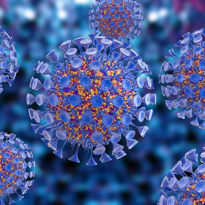
Literature - Publication Review
Applications of In Vivo bioluminescence imaging for SARS-CoV-2
Scientists continue to explore several options to treat SARS-CoV-2 infection with hundreds of therapeutics at various phases of...
Brochure
Biologics workflow solutions
Precision biologics are playing an increasingly powerful role as part of therapeutic strategies such as monoclonal antibodies...
SDS, COAs, manuals and more
Are you looking for technical documents related to the product? We have categorized them in dedicated sections below. Explore now or request your COA/TDS, SDS, or IFU/manual.
0 of 7 selected
Labeling Kits and Dyes
Part number:NEV11118
List price:USD 392.19/each
Your online price:Log in to view
Total list price:
USD 0
Part number:NEV12001
List price:USD 110.77/each
Your online price:Log in to view
Total list price:
USD 0
Lentiviral Particles
Part number:CLS960002
List price:USD 2,101.20/each
Your online price:Log in to view
Total list price:
USD 0
Probes
Part number:NEV10003
List price:USD 581.26/each
Your online price:Log in to view
Total list price:
USD 0
Part number:NEV10054EX
List price:USD 580.45/each
Your online price:Log in to view
Total list price:
USD 0
Part number:NEV10091
List price:USD 600.68/each
Your online price:Log in to view
Total list price:
USD 0
Part number:NEV10100
List price:USD 706.60/each
Your online price:Log in to view
Total list price:
USD 0
0 of 7 selected


How can we help you?
We are here to answer your questions.