Revvity Sites Globally
Select your location.
*e-commerce not available for this region.


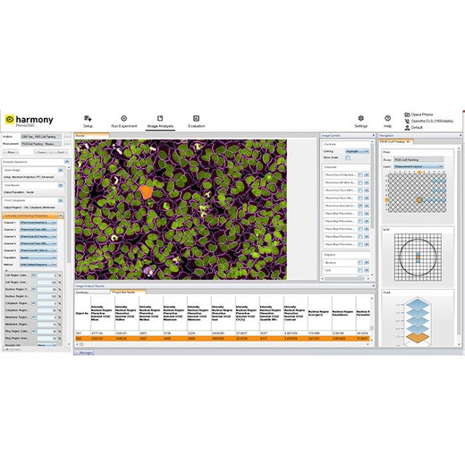
Harmony High-Content Imaging and Analysis Software
Harmony High-Content Imaging and Analysis Software

Harmony 1
Harmony High-Content Imaging and Analysis Software
Product variant
Unit Size: 1 each
Part #:
HH17000019
For research use only. Not for use in diagnostic procedures.
Product information
Overview
Designed for biologists, Harmony’s workflow-based interface makes the whole process of high-content analysis straightforward, even for new users with little microscopy or programming knowledge.
One high-content analysis software for all your applications
- Phenotypic Screening
- Get robust phenotypic fingerprints based on intensity, morphology, texture and fluorescence distribution parameters
- Easily train the software to recognize relevant phenotypes
- Quantify even subtle phenotypic differences
- Live Cell Imaging
- Follow phenotypic changes over time
- Accurately quantify label-free live cell images
- Track individual cells over time or analyze wound-healing assays
- Analyze cell signaling dynamics using e.g. FRET based assays
- Perform fast response assays such as calcium flux or cardiomyocyte beat rate with imaging frame rate of up to 100fps.
- Set-up "dispense and read" assays on the Opera Phenix Plus system with liquid handling module
- Enhance your live-cell assay capabilities through improved time-series flexibility
- 3D Cell Models
- Speed up 3D image acquisition by targeted imaging of microtissues
- Better understand your cell models by exploring them in the new 3D viewer and the XYZ viewer
- Measure morphologies and volumes in 3D, count nuclei within spheroids
- Enhance Maximum Intensity Projection Analysis using PlaneMap
- One software for image acquisition, 3D visualization and analysis
- Rare Cell Phenotypes
- Quickly capture your rare cell phenotypes with PreciScan™
- Pre-screen at low resolution and acquire objects of interest at high resolution
- Shorten your image acquisition times
- Reduce your data volume
- Routine Assays
- Utilize more than 30 ready-made-solutions for standard assays
- Ensure repeatability by running established protocols
- Optional module
- Phenologic.AI offers an efficient and reliable method for identifying cells and cellular nuclei within fluorescent and brightfield images by harnessing the power of pre-trained deep neural networks (DNNs).
One high-content analysis software for all users
- New Users
- Get quantification for your cell images
- Easy to learn – even for users with little microscopy or programming knowledge
- Get started with 30 ready-made solutions
- Logical workflow for easy control of all aspects of image acquisition and analysis
- Simple building blocks to assemble image analysis sequences (step-by-step)
- Expert Users
- Quantify difficult phenotypes with advanced texture and morphology readouts
- Utilize advanced building blocks to enable e.g. ratiometric FRET imaging
- Create “stitched” global images from multiple fields of view
- Examine samples at multiple scales – regions found in the global image can be used for analysis in the original images
- For Lab Managers
- Keep up with increasing research needs – analyze larger sample sizes
- Quickly train new users to set up the instrument and analyze their images
- Find images, metadata and results quickly via the integrated sortable database
- View and analyze data from any computer with installation of Harmony software
- Get application support by image analysis experts
Specifications
| Instrument Compatibility |
Opera
Opera Phenix Plus
Operetta CLS
|
|---|---|
| Unit Size |
1 each
|
Resources
Are you looking for resources, click on the resource type to explore further.
Brochure
3D cell culture workflow solutions
More than ever, researchers are turning to 3D cell cultures, microtissues and organoids to bridge the gap between 2D cell cultures...
Application Note
Analysis of mitochondrial dynamics in human iPSC-derived neurons using the Operetta CLS High-Content Analysis System
Mitochondrial dynamics are essential for energy conversion and neuron survival. Understanding changes in mitochondrial dynamics is...
Whitepaper
Artificial intelligence, machine learning and deep learning: applications in cellular imaging for improved drug discovery productivity
There has been a lot of buzz around artificial intelligence, machine learning and deep learning. Is the reality living up to the...
Application Note
Cytotoxicity studies on 3D primary liver microtissues
Liver toxicity remains one of the main reasons for drug failure in clinical trials. Improving preclinical toxicity testing is a...
Infographic
Deeper insights from your 3D cell model imaging
Researchers are increasingly looking to 3D cell models to bridge the translational gap between 2D cell cultures and in vivo...
Loading...


How can we help you?
We are here to answer your questions.