Revvity Sites Globally
Select your location.
*e-commerce not available for this region.

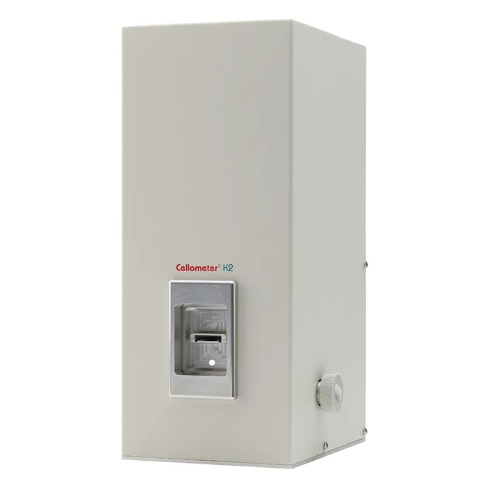
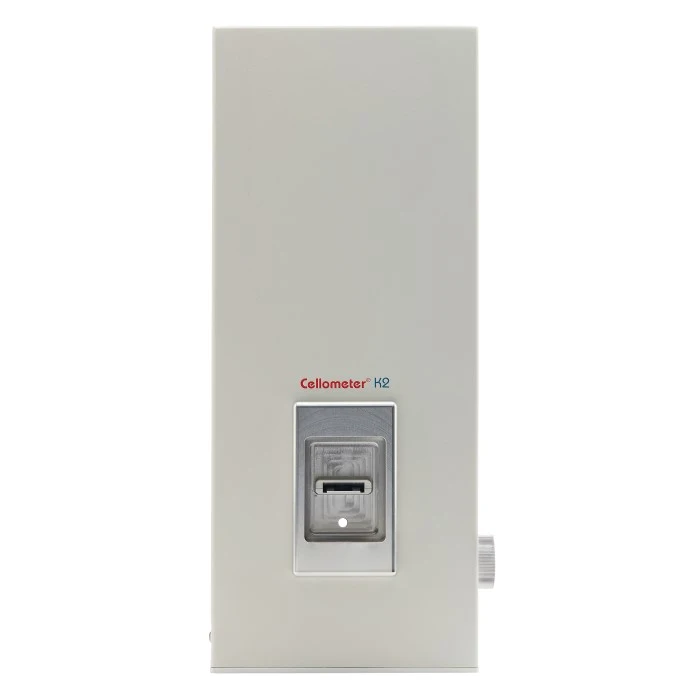
Cellometer K2 Fluorescent Cell Counter
For research use only. Not for use in diagnostic procedures.
Part #:
CMT-K2-MX-150
Imaging Modality:
Fluorescence, Brightfield

Cellometer K2 Fluorescent Cell Counter

cellometer-k2 700x700
Cellometer K2 Fluorescent Cell Counter
Product information
Overview
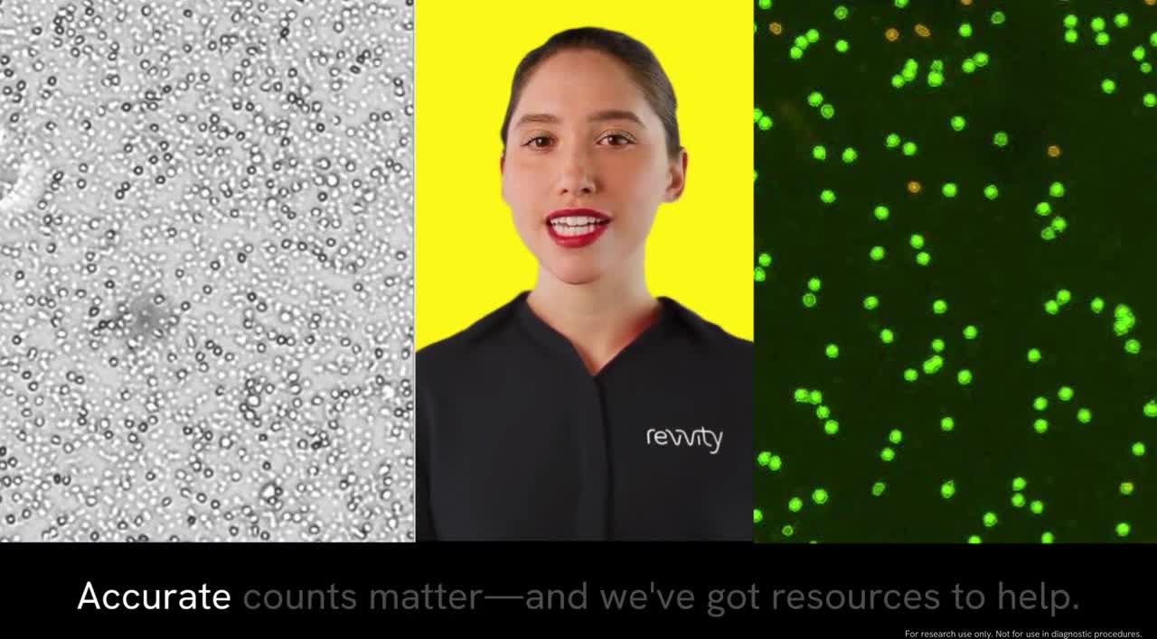
The Cellometer K2 fluorescent cell counter, powered by Matrix software, utilizes brightfield imaging and dual-fluorescence imaging to quickly and accurately identify and count individual cells. Cell count, concentration, diameter, and % viability are automatically calculated and reported.
The Cellometer K2 cell counter provides:
- Dual fluorescence and brightfield imaging – stain only nucleated cells for accurate count and viability information
- Fast results – count, size, concentration, and viability calculations in <60 seconds
- Analysis of complex samples – designed for analysis of complex and messy samples including whole blood, peripheral blood, cord blood, and bone marrow
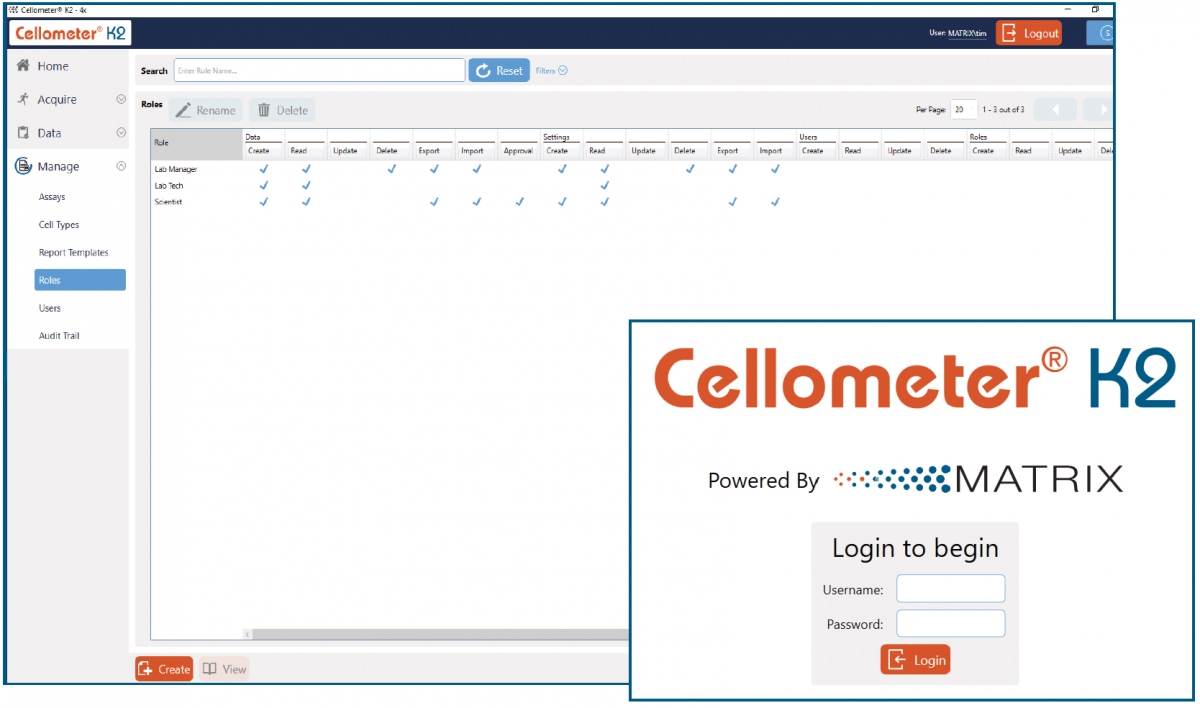
- 21 CFR Part 11 ready – optional add-on that includes an audit trail, user access control, and digital signature
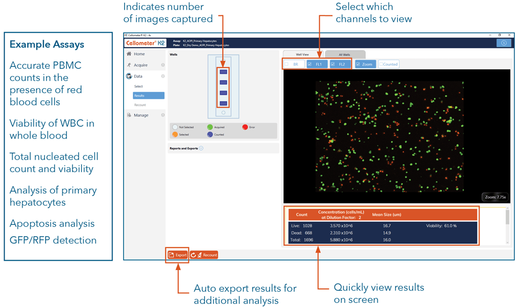
- Multiple fields of view – increased accuracy with the ability to capture one, four, or eight images per sample
- Built-in predefined assays – quickly analyze viability, apoptosis, and transfection efficiency
- Built-in cell types – includes saved parameters for over 400 cell types
- Small sample volume – only 10 µl of cell sample required
- Configurable reports – includes predefined reports with the ability to create new ones with graphs, images, charts, and tables
Additional product information
Count a variety of samples - from cell lines to primary samples
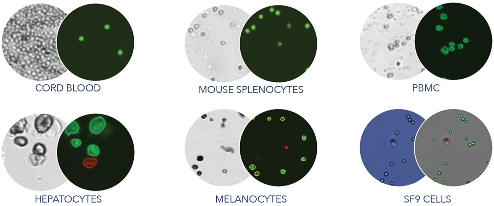
The Cellometer K2 can be configured to handle a variety of cell types, including primary cells, tumor digest, insect cells, cell lines, fragile cells, and more at low or high concentrations.
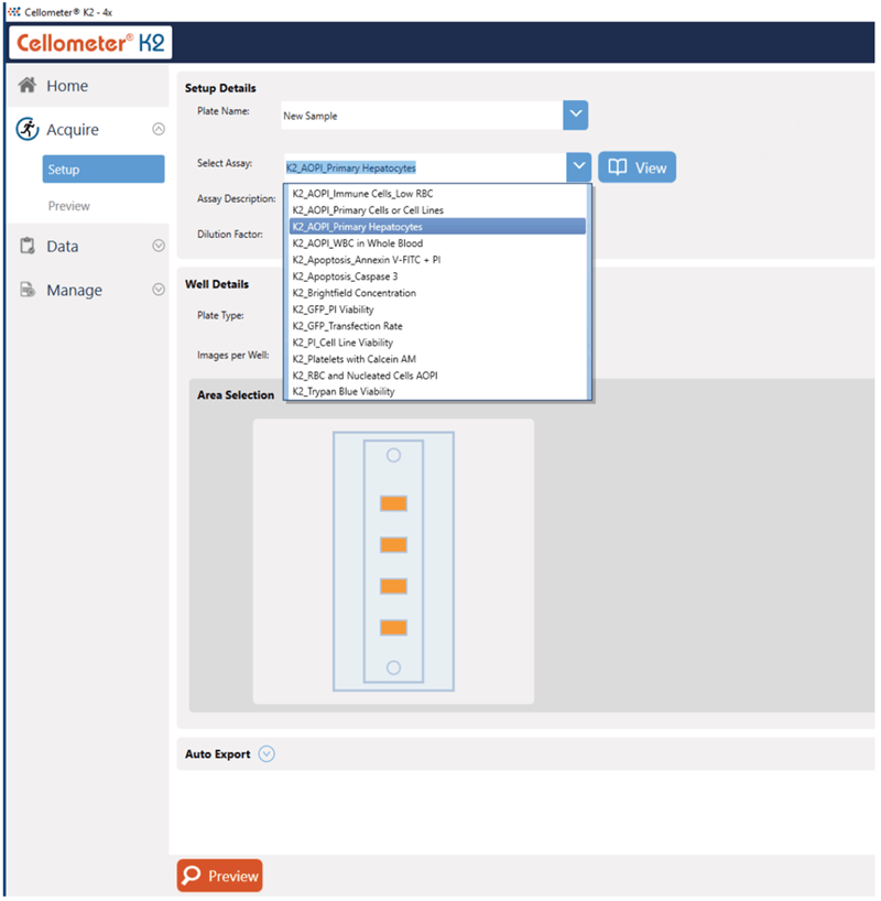
Predefined assays and cell types
Take the guesswork out of setting up your cell quantification experiments. The Cellometer K2 includes a library of commonly-used assays and cell types with predefined settings to help improve consistency of results. Conveniently build and modify assays and cell types to help match your specific experimental requirements.
Capture multiple fields of view
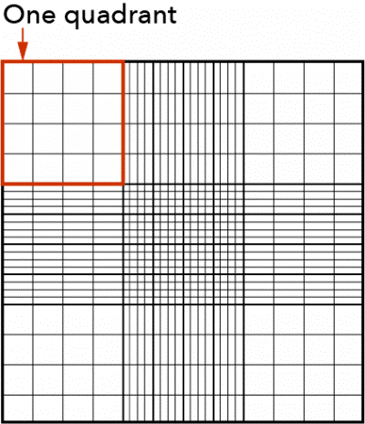
Capture one, four, or eight images per sample. The instrument default is set to four images, which is the equivalent of six quadrants on a hemocytometer. Eight images are equivalent to twelve quadrants on a hemocytometer. The ability to capture multiple images improves the cell counting dynamic range and reliability of results.
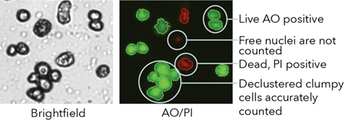
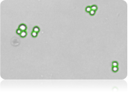
Dual-fluorescent staining for clumpy cells
The fluorescent image shows bright green AO-positive hepatocytes declustered by the Cellometer K2 algorithm. Red circled hepatocytes are PI-positive (dead) while free nuclei are not counted.
Predefined and customizable reports
Use default or customized reports based on the data and images you want to be included for your specific experimental needs. Automatically export images and data reports including CSV, Excel, Word, or PDF files. Perform statistical analysis for a wide range of parameters such as average, variance, min/max, and standard deviation of cell size.
21 CFR Part 11-ready
An optional module can be purchased to facilitate 21 CFR Part 11 compliance. The additional module comes with the following features:
- User login with passwords
- User assigned permissions
- Audit trail
- Error log files
- Electronic signatures
Live/dead nucleated cell counts using dual-fluorescence
Why dual-fluorescence?
Because brightfield cell counting does not differentiate nucleated from non-nucleated cells and Trypan blue staining is not as easy to detect as fluorescent staining, dual-color fluorescence is strongly recommended for reliable viability analysis for primary cells. The K2 is equipped with standard assays for dual-fluorescence analysis of a variety of cells stained with Acridine Orange and Propidium Iodide (AO/PI).
The AO/PI method
Acridine Orange (AO) is a nuclear staining (nucleic acid binding) dye permeable to both live and dead cells. It stains all nucleated cells to generate green fluorescence. Propidium Iodide (PI) can only enter dead cells with compromised membranes. It stains all dead nucleated cells to generate red fluorescence. Cells stained with both AO and PI fluoresce red due to quenching, so all live nucleated cells fluoresce green and all dead nucleated cells fluoresce red.
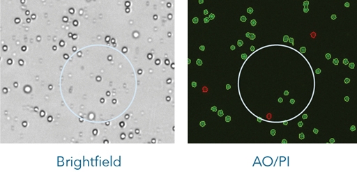
No interference from red blood cells, platelets, or debris
The dual-fluorescence AO/PI method utilizes nuclear staining dyes that bind to nucleic acids in the cell nucleus. Because most mature mammalian red blood cells do not contain nuclei, only live and dead mononuclear cells produce a fluorescent signal. There is no need to lyse red blood cells, saving time and eliminating an extra sample preparation step. Red blood cells, platelets, and debris are not counted in the fluorescent channels.
Analysis of clumpy and irregular-shaped cells
NCI-60 and clumpy MCF-7 cells
NCI-60 is a group of 59 human cancer cell lines (originally 60) developed by the National Cancer Institute (USA) for screening purposes.
- 57% of the NCI-60 cell lines are clumpy, contain debris, or display large variations in cell shape or size
- All 59 NCI-60 cell lines have been successfully validated on the Cellometer cell counter
Clumpy cells
Irregular-shaped cells

10x faster than manual counting
Counting 1 x 10⁶ cells takes approximately 5 minutes with a manual hemacytometer. Counting live and dead cells sometimes takes twice as long. The Cellometer K2 cell counter calculates cell count and concentration for live and dead cells and % viability in just 60 seconds.
Improve data reliability and consistency:
- Reduce judgment errors
- Reduce interference from RBCs
- Reduce recording & calculation errors
- Reduce counting time - run more experiments
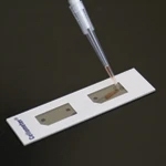
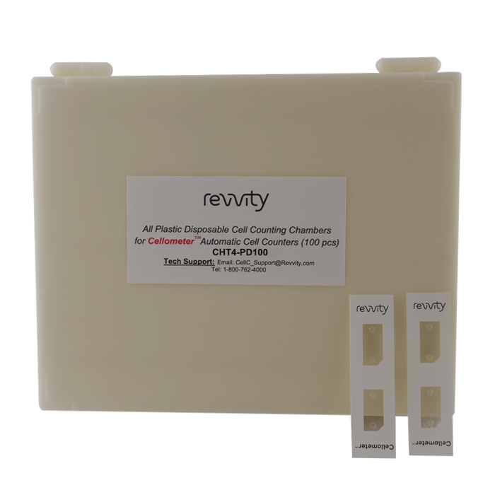
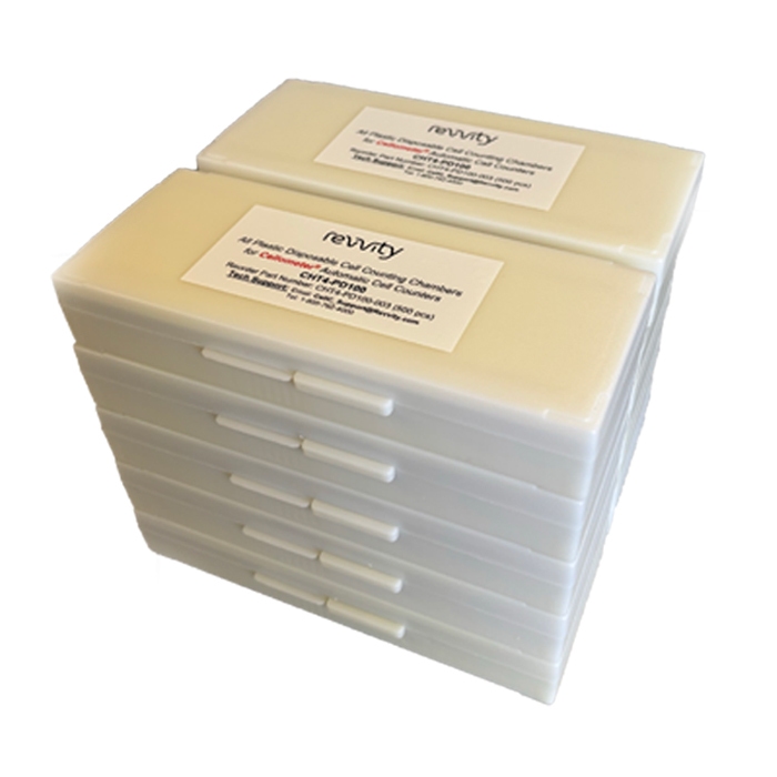
Imaging / counting chambers: no washing or contamination
We offer two different disposable counting slides, one with protective coverings on both sides and a ready-to-use option packed in microscope slide boxes. Disposable slides offer the advantages of time-saving, since no washing is required, reduced risk of contamination and reduced biohazard risk to users.
Specifications
| System includes |
|
|---|---|
| Available accessories |
|
| Imaging performance |
|
| Instrument specifications |
|
| PC / Laptop Minimum Requirements: (If purchasing Cellometer without PC laptop) |
|
| Available fluorescence optics modules |
|
Specifications
| Brand |
Cellometer
|
|---|---|
| Imaging Modality |
Fluorescence, Brightfield
|
| Unit Size |
1 each
|
Image gallery
Cellometer K2 Fluorescent Cell Counter
Cellometer K2 Fluorescent Cell Counter
Resources
Are you looking for resources, click on the resource type to explore further.
eBook
10 key factors to improve your cell counting results
Cell counting plays a crucial role in the development and manufacturing of cell and gene therapies as well as regenerative...
Brochure
Automated cell counting and image cytometry solutions brochure
With expertise and a pioneering spirit, Revvity boldly leads the way in cell imaging and analysis. As creators of advanced...
Flyer
Cellometer K2 fluorescent cell counter - designed for precise cell counting and fluorescent analysis
Related products
0 of 11 selected
AOPI Viability Reagents
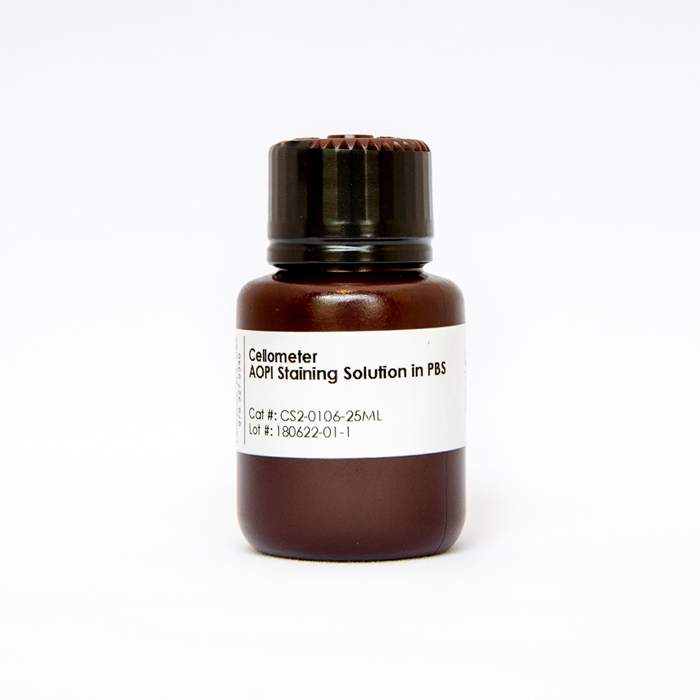
Part number:CS2-0106-25ML
List price:USD 577.16/each
Your online price:Log in to view
Total list price:
USD 0
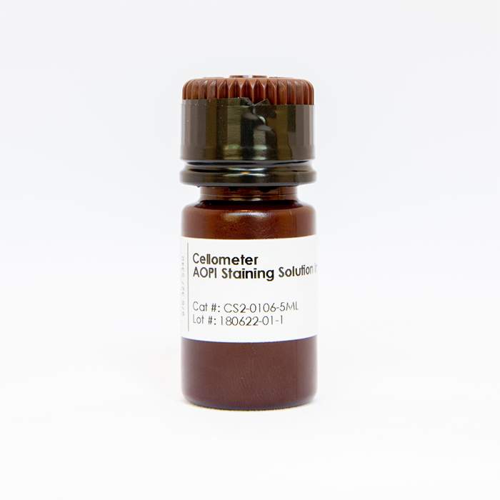
Part number:CS2-0106-5ML
List price:USD 128.87/each
Your online price:Log in to view
Total list price:
USD 0
Cell Counting Slides - PD100
Part number:CHT4-PD100-002
List price:USD 383.30/each
Your online price:Log in to view
Total list price:
USD 0
Part number:CHT4-PD100-003
List price:USD 1,732.50/each
Your online price:Log in to view
Total list price:
USD 0
Part number:CHT4-PD100-503
List price:USD 7,450.80/each
Your online price:Log in to view
Total list price:
USD 0
Cell Counting Slides - PD300
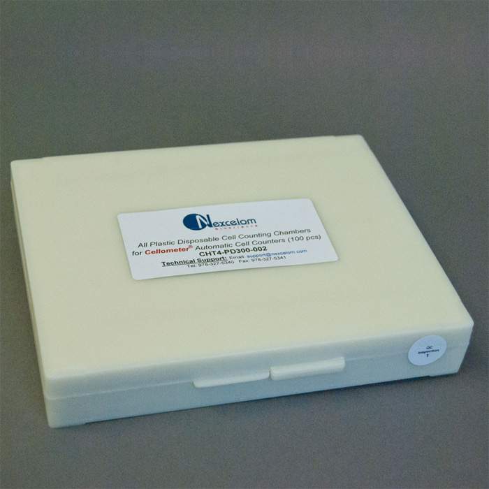
Part number:CHT4-PD300-002
List price:USD 480.23/each
Your online price:Log in to view
Total list price:
USD 0

Part number:CHT4-PD300-003
List price:USD 2,040.15/each
Your online price:Log in to view
Total list price:
USD 0
Cell Counting Slides - SD100

Part number:CHT4-SD100-002
List price:USD 236.82/each
Your online price:Log in to view
Total list price:
USD 0
Part number:CHT4-SD100-014
List price:USD 1,732.50/each
Your online price:Log in to view
Total list price:
USD 0
Part number:CHT4-SD100-314
List price:USD 4,975.95/each
Your online price:Log in to view
Total list price:
USD 0
Part number:CHT4-SD100-514
List price:USD 7,600.95/each
Your online price:Log in to view
Total list price:
USD 0
0 of 11 selected


Recently viewed

Part number:
INF02358
How can we help you?
We are here to answer your questions.