


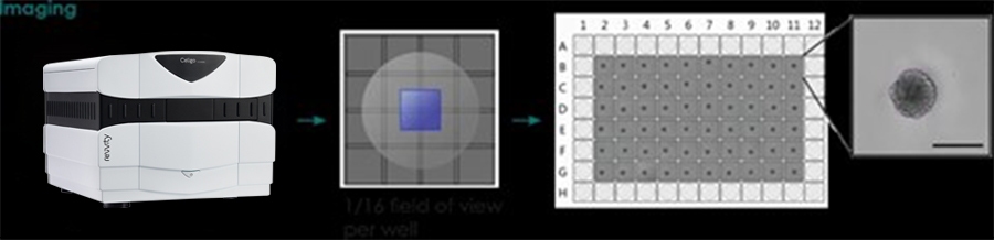
- Directly image tumor spheroids in various microwell formats
- Non-invasive brightfield imaging allows the user to image the same plate over multiple days
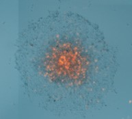
- Perform a two-color fluorescent viability assay
Introduction
The Celigo™ image cytometer has been developed to fully automate live cell analysis of tumorspheres. This automated morphometric analysis tool significantly reduces the time and effort needed to quantify key aspects of 3D spheres including size, growth, growth tracking over time and response to chemotherapeutics.
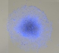
Measured Outgrowth from a HUVEC 3D Tumor Spheroid
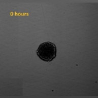
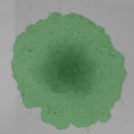
Spheroid Brightfield image
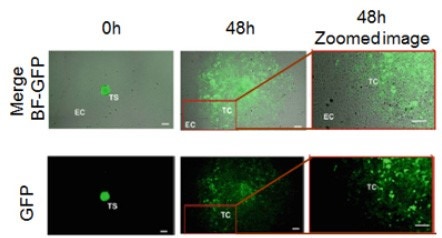
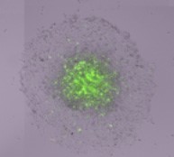
Monitoring cell outgrowth
from a GFP-labeled HUVEC spheroid
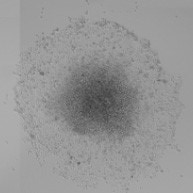
Spheroid Brightfield image
Spheroid fill view Brightfield image

For research use only. Not for use in diagnostic procedures.